Acoustic droplet vaporization is the focused ultrasound-mediated conversion of perfluorocarbon droplets into microbubbles. We are researching the use of this phenomenon in the induction of targeted occlusions in tumor vasculature. The goals are to enhance the efficacy of drug delivery and to "starve tumors to death" by cutting off their blood supply. We currently utilize the rat cremaster for intra-vital microscopy experiments and an ectopic hepatocellular carcinoma model in mice to test our proposed treatment. We are developing ultrasound-guided treatment methods to refine the spatial localization of the therapy.
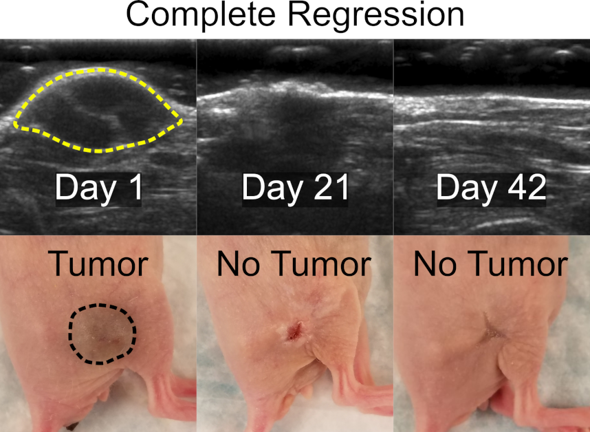
Gas embolotherapy is capable of achieving complete tumor regression in our mouse model.
Acoustic droplet vaporization-induced occlusion seen in the rat cremaster muscle.
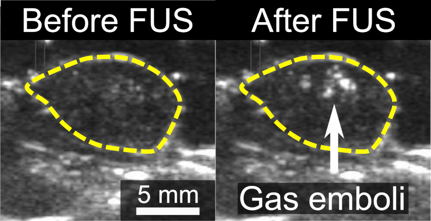
Ultrasound-guided treatment allows for direct visualization of gas emboli within a tumor.
Our Verasonics research ultrasound system provides us with fine control over acoustic transmit and receive operations, as well as easy access to raw data for custom processing. We have implemented a number of specialized imaging modalities, with an emphasis on tumor identfication, treatment planning, and treatment assessment. These include methods to image the mechanical properties of tissue (shear wave elastography) as well as tumor vasculature (super resolution vascular imaging) and relative hemoglobin oxygenation (photoacoustics).
Visualization of a propagating shear wave (left to right) in a phantom. These data can be used to determine local viscoelastic tissue properties.
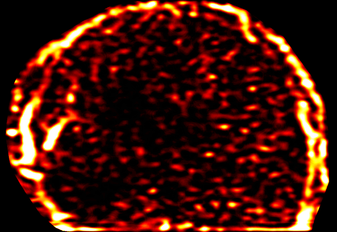
Super resolution image of tumor vasculature. Microbubble contrast agents and specialized image processing can be used to isolate vessel signal from tissue signal. Vessels with faster flow appear brighter.
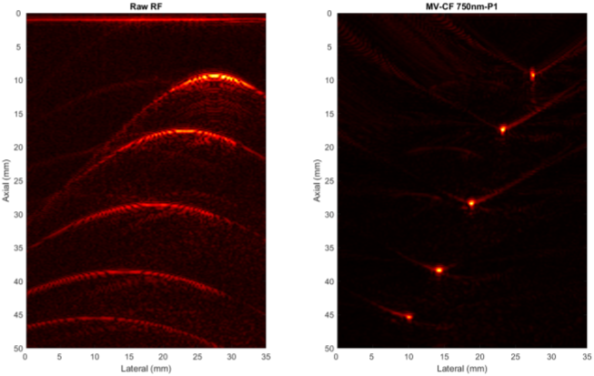
Easy access to raw data allows for custom beamforming of photoacoustic data (shown above), among other applications. In vivo, these imaging techniques can measure tissue oxygenation.
In this project, we perform simulations on nitric-oxide (NO) releasing catheters that combat bacterial infection and thrombosis. Emphasis is placed on simulating the NO concentration profile near the surface of the catheter and the NO flux out of the catheter. The simulations will help inform the design of the catheter to have a safe level of NO release without sacrificing the clinical benefits.
Simulation of the NO concentration profile in a dual-lumen catheter.
Lung Comets (B-lines) are imaging artifacts seen in clinical lung ultrasound. Excess lung comets indicate the presence of pulmonary edema. We focus on developing automatic quantification methods for lung ultrasound comets that improve the consistency of counting by applying image processing and machine learning methods. An accurate and consistent quantification of the lung comets may be useful in quantifying lung edema and aiding diagnosis.
Computer identified ultrasound comets in a B-mode cine loop (right, red lines).
Many therapeutic drugs can be toxic when administered systemically; the most common example is chemotherapy. Additionally, systemic administration is challenging for certain applications, particularly for delivering compounds to the brain. To circumvent these issues, we have fabricated custom droplets, capable of being loaded with an aqueous solution of a chosen drug, which will locally release the payload upon vaporization at a target site. Ongoing experiments include characterization of these droplets in vitro, particularly with respect to their ability to increase local drug uptake via mechanical disruption of vessels or cell membranes.
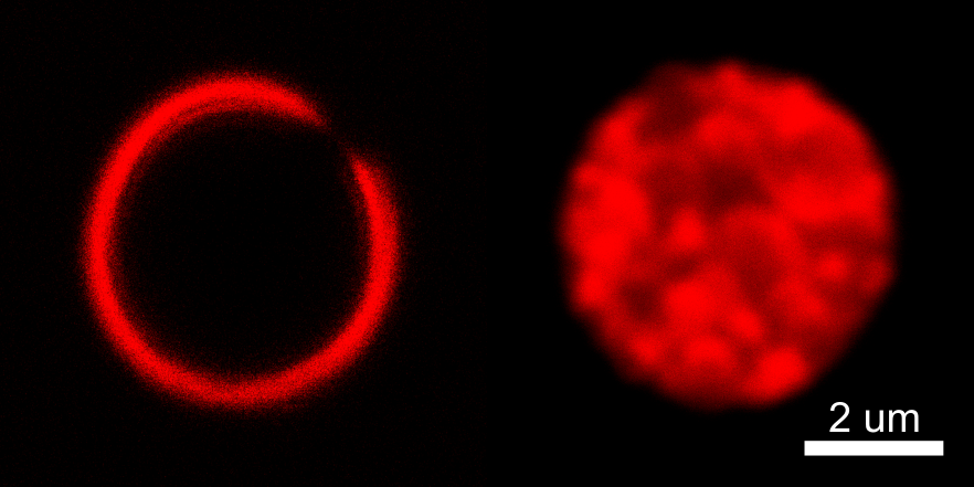
Droplets with doxorubicin, a highly cardiotoxic chemotherapeutic, loaded into either the shell (left) or the core (right).